mobilewounddocs.com

Are you a Long-Term Care Facility Seeking Superior Wound Care Services?

PROVIDING WOUND CARE TO NON-AMBULATORY PATIENTS AND THOSE WITHOUT ACCESS!

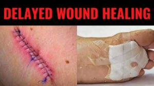
What is Delayed Wound Healing?

Daniel Davidson, MD, MBA, DBA, PHD Introduction: The physiological process of wound healing is intricate and includes several phases, such as tissue creation, remodeling, and inflammation. Most of the time, wounds move through these phases quickly, resulting in a successful closure and the preservation of tissue integrity. But occasionally, the healing process could take longer than expected, which could present difficulties for both patients and medical professionals. Numerous variables, from underlying medical issues to environmental circumstances, can cause delayed wound healing. We examine the reasons, signs, and available treatments for delayed wound healing in this article. What is Delayed Wound Healing? A condition known as “delayed wound healing” occurs when the body’s normal healing process—which normally involves mending damaged tissue—takes longer than anticipated or does not proceed through the phases in a reasonable amount of time. The biological process of wound healing is intricate and includes tissue creation, remodeling, and inflammation. In most cases, wounds close successfully and tissue integrity is restored as they move through these phases in a timely manner. Delays can occur from a variety of circumstances interfering with the natural healing process. These could include underlying medical disorders that affect blood flow, weaken the immune system, or obstruct the supply of vital nutrients needed for tissue regeneration, such diabetes, vascular disease, or malnutrition. Another frequent reason for delayed wound healing is infection, which is brought on by microbiological invasion, which can also worsen tissue regeneration, lengthen inflammation, and raise the risk of consequences. Causes of Delayed Wound Healing: A number of things can cause delayed wound healing, and all of them can make it more difficult for the body to restore injured tissue. These are a few typical reasons: Underlying Health Conditions: Wound healing can be severely hampered by long-term conditions such diabetes, vascular disease, autoimmune disorders, and malnutrition. These ailments may weaken the immune system, impair blood flow to the wound site, or obstruct vital nutrients needed for tissue healing. Infection: Infected wounds have a higher probability of taking longer to heal. Infections with bacteria, fungi, or viruses can exacerbate tissue regeneration, prolong inflammation, and raise the possibility of consequences including sepsis or abscess formation. Inadequate Blood Supply: To support tissue regeneration and healing at the wound site, enough blood flow is necessary to deliver nutrients and oxygen. Healing can be hampered by conditions that impair blood supply, such as venous insufficiency or peripheral artery disease. Drugs: A number of drugs have the potential to obstruct the body’s natural healing processes, delaying the healing of wounds. Corticosteroids, immunosuppressants, and nonsteroidal anti-inflammatory medications (NSAIDs) are a few examples. Smoking:  By narrowing blood vessels, lowering oxygen flow to tissues, and compromising immunological function, tobacco use can hinder the healing of wounds. Smokers are more likely to experience problems after surgery or an injury, including delayed wound healing. Advanced Age:  Aging is linked to alterations in the structure of the skin, a decrease in the generation of collagen, and a compromise in immunological function, all of which can cause delays in the healing of wounds. Nutritional Deficiencies: Encouraging the body’s healing processes requires a healthy diet. Wound healing can be hampered by deficiencies in protein, vitamins (especially C and A), and minerals (such iron and zinc). Obesity: Carrying too much weight around can put strain on wounds, obstruct blood flow, and raise the possibility of problems like infection. Poor outcomes after surgery or injury and delayed wound healing are linked to obesity. Poor Wound Care: Inadequate wound care can slow down the healing process and raise the risk of complications. This includes inappropriate cleaning, dressing selection, and failure to shield the site from additional trauma or infection. Symptoms of Delayed Wound Healing: Persistent Redness: Prolonged redness or inflammation in the vicinity of the wound may be a sign of continued inflammation and poor healing. Swelling: Prolonged edema or swelling close to the wound site may be a sign of a delayed healing process, which is frequently brought on by fluid buildup and poor lymphatic drainage. Warmth: If the skin around the incision feels warmer than the surrounding skin, there may be persistent inflammation and insufficient healing taking place. Pain: Continued or worsening pain at the site of the wound, especially during the initial phases of healing, may be a sign of underlying problems such tissue damage, nerve involvement, or infection. Increased Drainage: Extended periods of significant pus, blood, or clear fluid drainage from wounds may be a sign of infection or delayed healing. Reopening of the Wound: After first closure, wounds that frequently deteriorate or reopen may be a sign of underlying problems with tissue regeneration and healing. Development of Granulation Tissue: A symptom of the healing process, granulation tissue takes the form of pink or red tissue in the wound bed. On the other hand, extensive or protracted granulation tissue production could be a sign of postponed healing. Symptoms throughout the system:  Systemic symptoms like fever, chills, weariness, and malaise can appear in severe cases of delayed wound healing, especially if an infection is present. Treatment of Delayed Wound Healing: Wound Debridement: Objective: By minimizing the chance of infection and establishing a clean wound bed, removing dead or necrotic tissue from the wound site aids in the healing process. Techniques: There are a number of ways to accomplish debridement, including as mechanical, enzymatic, autolytic, or surgical approaches. Procedure: Depending on the features and severity of the wound, medical professionals will carefully remove non-viable tissue using sharp instruments, specialty dressings, or topical treatments. Infection Control: Objective: In order to lower inflammation, avoid systemic problems, and encourage tissue regeneration, it is essential to treat underlying infections. Method: Depending on the kind of infection present and the outcomes of culture and sensitivity testing, medical professionals may recommend antimicrobial therapy, such as antibiotics, antifungals, or antivirals. Monitoring: Treatment choices are based on a routine evaluation of the site for indicators of infection, such as elevated redness, edema, temperature, or purulent drainage. Optimizing Nutrition: Objective: By giving the body the vital

Read More »

What is Decubitus Ulcer and how is it Formed?

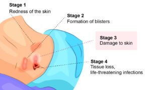
Daniel Davidson, MD, MBA, DBA, PHD Introduction: Decubitus ulcers, sometimes called bedsores or pressure ulcers, these are a frequent medical ailment that can be prevented by applying pressure to the skin. The condition arises when pressure is applied to certain areas of the body, reducing blood flow to those areas. As a result of continuous pressure on particular body parts, these ulcers usually develop in people who are confined to a bed or chair for extended periods of time. Formation of Decubitus Ulcers: Long-term pressure on the skin causes reduced blood flow to the affected area, which can result in decubitus ulcers. A prolonged period of pressure applied to the skin may harm the tissue beneath it. Tissue damage and ultimately the development of an ulcer are caused by the absence of blood flow, which deprives the tissue of nutrients and oxygen. Pressure: Pressure on the skin is the main factor contributing to decubitus ulcers. Blood flow is restricted to a certain place of the body when pressure is applied there for a prolonged amount of time. Ischemia: Also referred to as restricted blood flow, ischemia causes the affected tissue to lose vital nutrients and oxygen. Consequently, the tissue starts to die, causing harm to other tissues and ultimately the development of an ulcer. Friction and Shear: Besides pressure, decubitus ulcers can also develop as a result of friction and shear. Shear happens when tissue layers move against one another, whereas friction happens when the skin brushes against a surface. These pressures have the potential to further deteriorate the skin and underlying tissue, increasing the risk of ulcer development. Skin Breakdown: The skin and underlying tissue sustain damage as a result of continuous pressure, friction, and shear. A pressure injury may first show signs of redness or discoloration of the skin. An open sore or blister may develop if the pressure continues to build up on the epidermis, the skin’s outermost layer, and a portion of the dermis, the skin’s underlying layer. Ulcer Formation: Should the pressure remain unchanged, the ulcer may go through the following stages: Stage 1: Only the skin’s outermost layer is affected by the superficial ulcer. To the touch, the skin could appear heated, discolored, or red. Stage 2: An open sore or blister is formed as the ulcer spreads into the skin’s deeper layers. Stage 3: The tissue under the skin is harmed when the ulcer spreads into the subcutaneous layer. Stage 4: The worst stage, in which the ulcer penetrates deeply into the tissue and affects the bone, muscles, and tendons. Risk Factors: The following are some of the variables that can raise the risk of decubitus ulcers: Immobility: Those who are confined to a wheelchair or are bedridden are more vulnerable. Reduced mobility: Individuals who have restricted movement, such as those recuperating from injuries or surgeries, are also vulnerable. Inadequate nutrition: The body’s capacity to heal injured skin may be hampered by a diet deficient in important nutrients. Incontinence: Skin breakdown is more likely when there is an excessive amount of moisture in the urine or feces. Age:  Because their skin is thinner and their mobility is limited, older persons are more likely to get decubitus ulcers. Prevention and Treatment: Avoidance: Frequent Position Changes: Changing postures on a frequent basis is one of the best strategies to avoid decubitus ulcers. Those who are confined to a wheelchair or bed can reduce pressure on sensitive body parts by switching positions every two hours. Utilization of Support Surfaces: Pressure ulcer risk can be decreased and pressure can be distributed more uniformly with the use of specialized mattresses, cushions, and padding. These support surfaces lessen strain on the body’s susceptible parts, such as bony prominences. Sustaining Proper Nutrition:  In order to keep skin healthy and stop pressure ulcers from developing, a balanced, healthful diet is crucial. Sufficient consumption of protein, vitamins, and minerals is especially crucial for fostering tissue regeneration and averting skin deterioration. Skin Care: The prevention of decubitus ulcers depends on maintaining clean, dry skin. Regular skin hydration can reduce the possibility of dryness and cracking, which raises the danger of skin damage. Maintaining dry and clean skin is especially crucial when incontinence is a problem. Preventing Friction and Shear:  These two factors raise the possibility of pressure ulcer development and skin deterioration. Those who are bedridden or wheelchair-bound should be moved or repositioned carefully to prevent dragging or tugging the skin. Treatment: Relieving Pressure: Relieving pressure on the affected area is the initial step in treating decubitus ulcers. This can entail often shifting positions, applying support surfaces like orthopedic mattresses or pillows, and avoiding applying pressure to the afflicted area. Wound Care: To encourage healing and stave off infection, the wound must be kept clean and moist. To encourage healing, wounds should be cleansed with a mild cleanser and dressed with moisture. Debridement: To remove dead or necrotic tissue from a wound and encourage healing, debridement may be required in some situations. There are a number of techniques for debridement, including as autolytic, enzymatic, and sharp debridement. Bandages and Dressings:  Covering a wound with bandages or dressings can aid in healing and shield it from additional harm. Depending on the kind and severity of the wound, there are several types of dressings available, such as foam dressings, alginate dressings, and hydrocolloid dressings. Pain control:  Because decubitus ulcers can cause excruciating pain, it’s critical to provide patients with appropriate pain control in order to enhance their comfort and overall health. To treat pain brought on by decubitus ulcers, physicians may prescribe painkillers in addition to non-pharmacological therapies including wound care and posture. Conclusion: A common, but potentially dangerous, medical disease known as decubitus ulcers can arise when pressure on the skin inhibits blood flow to particular parts of the body. People can lessen their chance of getting these excruciating and possibly fatal sores by learning the causes and risk factors of decubitus ulcers and taking preventative approaches. For the purpose of avoiding problems and

Read More »

What is Contaminated Wound, and How does that Delay Healing? Why does Contamination Delay Healing? How to Treat it?

Daniel Davidson, MD, MBA, DBA, PHD Introduction: Bacteria, fungi, and viruses can contaminate wounds, which can range in severity from little cuts to serious traumas. A wound gets contaminated by foreign bacteria, which raises the risk of infection and slows down the healing process. We examine the idea of contaminated wounds, how contamination hampers healing, and practical treatment options in this article. What is a Contaminated Wound? Any injury or break in the skin that has come into contact with potentially dangerous germs, foreign objects, or substances is referred to as a contaminated wound. Numerous things can contaminate an area, such as coming into contact with dirt, garbage, bodily fluids, or pathogen-carrying objects. Microorganisms can cause infection and obstruct the body’s natural healing process when they infiltrate a wound. Lacerations, abrasions, puncture wounds, and surgical incisions exposed to non-sterile surroundings are common instances of contaminated wounds. Thorough wound assessment, decontamination, infection control, and the use of the right wound care therapies to encourage healing and avert complications are all necessary for the effective management of contaminated wounds. How Contamination Delays Healing: Pathogen Introduction: When a wound is contaminated, dangerous microorganisms like bacteria, fungus, and viruses are brought in. Infection may result from the proliferation of these organisms in the wound environment. An inflammatory response brought on by infection restricts the body’s natural healing process and expands its inflammatory phase. Inflammatory Response: In order to fight the invasive microorganisms, the body’s immune system releases inflammatory mediators in response to a contaminated wound. Though some degree of inflammation is necessary for the healing process, too much or too much of it can harm healthy tissue and prevent wounds from healing. Tissue Destruction: When a wound is exposed to foreign objects or substances that are poisonous to cells, contamination can cause tissue damage and necrosis. Destruction of tissue inhibits the growth of new tissue and produces an environment that is not conducive to recovery. Reduced Angiogenesis: Getting oxygen and nutrients to the wound site requires Angiogenesis, the growth of new blood vessels. Wound contaminants can hinder angiogenesis, which lowers blood flow and hinders tissue repair. Delayed Epithelialization: In order for a wound to close, epithelial cells must migrate and proliferate in order to cover the wound surface. By obstructing epithelial cell migration and preventing the creation of a new epithelial layer, contamination can postpone the process of epithelialization. Increased Complication Risk: Contaminated wounds are more likely to experience consequences like cellulitis, abscess formation, and sepsis, a systemic infection. These issues impede the healing process even more and can call for more drastic measures to address. Treatment of Contaminated Wounds: Wound Irrigation:  Objective: By flushing the wound with a sterile solution, debris, impurities, and pollutants are removed, lowering the risk of infection and the microbial burden. Method: To completely clean the wound, use an irrigation bottle or syringe to gently stream sterile saline solution or an antibacterial agent. Make sure there is enough pressure applied to remove material without endangering surrounding tissue. Debridement:  Objective: By establishing a clean, healthy wound bed, removing contaminated debris, foreign objects, and necrotic tissue from the wound site helps the wound heal. Techniques: Debridement can be accomplished by a number of methods, such as autolytic (moist dressings), enzymatic (topical enzymes), mechanical (wet-to-dry dressings), or surgical (sharp). Based on the patient’s tolerance and the features of the wound, choose the best technique. Topical Antimicrobial medications: Inhibiting microbial development, preventing infection, and accelerating healing are the goals of applying antimicrobial medications topically to wounds. Options: Take into account applying topical antimicrobial treatments including antimicrobial ointments, iodine solutions, or silver dressings. Based on the patient’s preferences, microbiological susceptibility, and wound characteristics, select the right agent. The purpose of dressings and wound covers ,Using the right dressings and coverings for the wound aids in preserving a moist environment, absorbing extra fluid, shielding the wound from more infection, and fostering tissue regeneration. Choices: Select from a range of dressings, such as films, foams, hydrocolloids, or alginates, depending on the size, depth, exudate level, and other parameters of the wound as well as the intended results. Systemic Antibiotics: Indication:  To remove pathogens and prevent complications, systemic antibiotic therapy may be required in cases with established infection or high risk of infection. Prescription: Whenever possible, prescribe antibiotics based on wound culture and sensitivity findings. Select broad-spectrum antibiotics to combat common wound bacteria, and modify treatment as necessary in response to the patient’s clinical condition. Pain Management: Things to Think about Intense discomfort can occur from contaminated wounds, particularly during debridement and dressing changes. Use efficient pain management techniques to guarantee patient comfort and treatment compliance.Choices: To reduce pain and suffering, use analgesic drugs, topical treatments (like lidocaine gel), or non-pharmacological therapies (such relaxation techniques and distraction tactics). Conclusion: Significant obstacles to the healing process arise from contaminated wounds, which raise the risk of infection and slow down tissue recovery. Comprehending the methods via which pollution obstructs healing is essential to putting treatment plans into action. Healthcare practitioners can maximize the healing results of contaminated wounds and reduce the risk of complications by utilizing appropriate wound care interventions, decontamination techniques, infection control measures, and thorough wound assessment. Timely wound healing and tissue integrity restoration depend on the early identification and timely control of contamination.

Read More »

What is a Suprapubic Tube and how is it Changed at the Bedside by a Physician?

Daniel Davidson, MD, MBA, DBA, PHD Introduction: For those who are unable to empty their bladders naturally through the urethral route, suprapubic tubes (SPTs) are essential passageways for urine drainage. Urinary diversion can now be reliably achieved with this minimally invasive surgical operation, which entails a small abdominal wall incision leading directly into the bladder to install a tube. A doctor’s bedside suprapubic tube change is a delicate process that demands accuracy, dexterity, and close attention to detail. We explore the nuances of suprapubic tubes, their purpose, and how to change them at the bedside in this article. What is a Suprapubic Tube? A medical device called a suprapubic tube (SPT) is used to drain urine when a person is unable to use the natural urethral pathway to empty their bladder. Through a tiny incision made in the lower abdomen, usually above the pubic bone, a hollow catheter is surgically inserted into the bladder. Using this other path, urine can constantly drain from the bladder and collect outside the body in a bag. When urinary retention arises from a variety of medical disorders, including neurogenic bladder dysfunction, urinary blockage, pelvic trauma, or bladder dysfunction following surgery, suprapubic tubes are frequently employed. They are also used in situations where traditional urethral catheterization is risky or impractical, including in strictures or urethral injuries. A qualified healthcare provider, such as a surgeon or urologist, will normally place a suprapubic tube in a clinical setting while under local or general anesthesia. During the surgery, a little incision in the lower abdomen wall is made, and the catheter is then advanced through it into the bladder. To keep the catheter from coming loose, it is usually fastened in place with sutures or a retention balloon. Procedure for Changing a Suprapubic Tube: In order to preserve urine drainage, changing a suprapubic tube is a delicate technique that requires replacing the old catheter with a new one. A medical professional or other qualified healthcare provider usually does this procedure at the patient’s bedside. A detailed tutorial explaining how to change a suprapubic tube can be found below: Assemble Materials:Sterile substitution suprapubic catheter (e.g., Foley catheter) of the proper size and kindsterile gloves antiseptic solution (such as povidone-iodine or chlorhexidine)draperies that are sterileNeedleslubricant gellocal anesthetic, if appropriateSecuring tool (such as a suture or retention balloon) Get the patient ready:Assume a comfortable supine or semi-recumbent position for the patient on the bed.Make sure there is enough light, and reveal the patient’s abdomen. To reduce nervousness, reassure the patient and go over the process with them. Wash and sanitize:Using mild circular motions, cleanse the area surrounding the current suprapubic tube insertion site with an antiseptic solution.Before continuing, let the antiseptic solution entirely dry. Anesthesia, if necessary:To decrease discomfort during the treatment and to numb the insertion site, use local anesthetic.Using a sterilized syringe and needle, inject a tiny quantity of lidocaine or another local anesthetic into the surrounding tissue.Await the anesthesia’s onset before moving forward. Removal of the tube:Remove the suture or adhesive dressing that is holding the existing suprapubic tube in place with caution.To extract the tube from the bladder, use mild traction and make sure to proceed slowly and deliberately to prevent damaging the surrounding tissue. New Tube Insertion:Apply a water-soluble lubricating lubricant to the tip of the sterile replacement suprapubic catheter using sterile technique.Applying sustained, gentle pressure, insert the catheter into the bladder through the site of the existing incision. Move the catheter slowly and cautiously, using tactile input and, if possible, fluoroscopic imaging as your guides.Make sure the catheter tip is inside the bladder and that the balloon (if any) is inflated in accordance with the instructions provided by the manufacturer to keep it there. Keeping the Tube Secure:To stop displacement, connect the new suprapubic tube to the skin with a suitable anchoring tool, like a suture or retention balloon.Make sure the tube is firmly in place and does not put too much strain on the tissue around it. Evaluation and Record-Keeping:Examine the suprapubic tube’s position and functionality to ensure that it is functioning properly and that enough pee is being drained from the bladder.After the procedure, keep an eye out for any indications of bleeding, discomfort, or problems in the patient. Keep track of all the procedure’s specifics, such as the kind and size of catheter used, any problems that arose, and the patient’s reaction. Conclusion: When it comes to facilitating urine drainage for people with different urological disorders, suprapubic tubes are essential. A suprapubic tube change at the patient’s bedside is a specialist treatment that needs strict adherence to sterile procedures and close attention to detail. Physicians can safely and successfully perform this treatment, guaranteeing the best possible patient outcomes and comfort, by according to established protocols and guidelines. Suprapubic tube management improves patient mobility, quality of life, and general well-being in addition to making urine drainage easier.

Read More »

What are the Surgical and Non-Surgical Modalities Utilized in the Management of Various Stages of Decubitus Ulcers?

Daniel Davidson, MD, MBA, DBA, PHD Introduction: Decubitus ulcers—also referred to as bedsores or pressure ulcers—pose a serious risk to patient safety and quality of life, making them a major healthcare concern. Decubitus ulcer treatment necessitates a multimodal strategy, with potential surgical and non-surgical interventions based on the ulcer’s particular stage. This article examines the range of surgical and non-surgical procedures used to treat decubitus ulcers at different phases of the condition. Stage-Based Management: The method of treating decubitus ulcers according to the degree of the wound—which is divided into stages that range from Stage I to Stage IV—is known as “stage-based management.” To encourage healing and stop more issues, each stage denotes a distinct degree of tissue involvement and calls for particular therapies. Stage I: Stage I ulcers are superficial sores that impact the skin’s outermost layer. The skin doesn’t break even if it looks red.Management and oversight: Put prevention first: By using pressure-relieving surfaces and relocating the damaged area, you can stop more harm from occurring.Preserve the integrity of your skin:  Prevent the skin from going to deeper stages by keeping it hydrated, free of friction, and clean. Stage II: Stage II ulcers are characterized by a partial thickness loss of epidermal integrity that penetrates into the dermis. The injury could manifest as a shallow crater, blister, or abrasion.Supervisory: Wound care:  To keep the wound moist and encourage healing, use the proper dressings. In addition to absorbing extra fluid, dressings should shield the wound from additional harm.Offload pressure: To stop additional tissue injury and promote recovery, keep up the pressure relief measures. Stage III: Stage III ulcers are characterized by a complete loss of epidermal integrity that extends into the subcutaneous region. The incision might resemble a large crater with visible fat.Supervisory: Debridement of the wound: In order to encourage healing and guard against infection, remove any necrotic or non-viable tissue from the ulcer site.Control of infections: Keep an eye out for symptoms, and if necessary, start the right antibiotic treatment.Offload pressure: To reduce pressure at the ulcer site, keep applying pressure relief techniques and make use of specific support surfaces. Stage IV: The most severe type of ulcers, known as stage IVs, entail full-thickness tissue loss that spreads into the muscle, bone, or supporting structures. A deep crater with exposed muscle, bone, or tendon may be the appearance of the wound.Surgical intervention: To encourage wound closure and tissue regeneration, take into account surgical methods such flap reconstruction, skin grafting, or tissue transfer operations.Wound vacuum therapy: By eliminating extra fluid and encouraging the growth of granulation tissue, negative pressure wound therapy (NPWT) helps to accelerate wound healing.Intensive wound care: To avoid problems and encourage healing, give careful wound care and infection control measures. Surgical Modalities: Surgical techniques are essential interventions in the management of decubitus ulcers, especially when the ulcers are progressed and non-surgical treatments may not be adequate. The goals of these surgical methods are to stop additional problems, seal the ulcer site, and encourage wound healing. Now let’s examine the many surgical techniques frequently applied to the treatment of decubitus ulcers: Rebuilding the Flaps:During a surgical process called flap reconstruction, healthy tissue from nearby parts of the body, such as skin, subcutaneous fat, and occasionally muscle, is transferred to cover the ulcer site. This procedure is especially helpful for large, complicated, or deep ulcers where conventional wound closure techniques might not be sufficient. Benefits:Gives the ulcer location a strong, well-vascularized tissue covering.Contributes to the healing of wounds by supplying healthy tissue with an unbroken blood flow.Lowers the chance of a wound healing poorly and returning. Flap Types: Rotational Flaps: To cover the ulcer location, tissue is rotated in from surrounding areas. Because of their versatility, rotational flaps can be made to fit the exact size and form of the wound.Advancement Flaps: To cover the ulcer, tissue is moved forward from nearby regions. To achieve tension-free closure and close larger faults, advancement flaps can be helpful. Skin Grafting:The goal of skin grafting is to encourage tissue regeneration and wound closure by transplanting healthy skin from a donor site to the ulcer site. Depending on how deeply tissue is taken, skin grafts can be divided into full-thickness or split-thickness categories. Benefits:Offers a comparatively easy and efficient way to seal wounds.Can be taken from a variety of donor locations, including as the patient’s own body (autograft), cadaveric sources (allograft), or artificial materials (xenograft).Encourages the healing of wounds and re-epithelialization. Skin Graft Types: Split-Thickness Skin Grafts: These skin grafts are made up of some dermis and some epidermis. When there is a enough supply of donor skin, they are frequently used for bigger wounds.Full-Thickness Skin Grafts:  The complete dermis and epidermis are grafted in these cases. They work well for smaller, shallower wounds and yield superior esthetic results. Procedures for Tissue Transfer: In order to encourage wound healing and tissue regeneration, vascularized tissue, such as muscle or fasciocutaneous flaps, is transplanted to the ulcer site during tissue transfer procedures. When there is underlying tissue loss in deep or widespread ulcers, these methods are especially helpful. Benefits:Gives densely vascularized tissue a steady blood supply, which encourages quick healing and lowers the chance of graft failure.Able to treat intricate wound flaws that expose bone or muscle.Lowers the chance of recurrence and provides long-lasting coverage. Non-Surgical Modalities: When it comes to the treatment of decubitus ulcers, non-surgical techniques are quite important, especially in the initial phases of wound development. Without requiring intrusive procedures, these therapies aim to stop more tissue damage, encourage wound healing, and lower the risk of complications. Pressure release: Pressure release is essential for stopping decubitus ulcers from getting worse. Blood flow to the injured tissues is enhanced by lessening pressure on sensitive parts of the body, such as bony prominences, which promotes healing and stops additional harm. Repositioning the patient on a regular basis, using cushions or mattresses that relieve pressure points, and maximizing patient mobility are all strategies for pressure reduction.Skin Maintenance: Maintaining skin integrity and halting

Read More »

What are the Legal Implications of the Formation of Decubitus Ulcers in Chronically Non-Ambulatory Patients Treated in Various Long Term Facilities?

Daniel Davidson, MD, MBA, DBA, PHD Introduction: Decubitus ulcers, sometimes referred to as bedsores or pressure ulcers, pose legal as well as medical difficulties, especially in the context of long-term care institutions where patients who are chronically non-ambulatory are frequently treated. In order to keep residents safe and healthy, these institutions have a responsibility to prevent decubitus ulcers from forming and to properly care for any sores that already exist. Healthcare professionals, workers, and facilities may face legal repercussions if these obligations are not fulfilled. This article examines the legal ramifications of decubitus ulcer development in patients receiving treatment in different long-term care institutions who are chronically non-ambulatory. Duty of Care: A legal notion known as the “duty of care” serves as the foundation for an organization’s or professional’s responsibility to take reasonable precautions to prevent injury to others. The term “duty of care” refers to the obligation placed on healthcare organizations and long-term care facilities to provide a level of care that guarantees the security, welfare, and best interests of their patients or residents. Standard of Care: Professional guidelines, industry conventions, and legal requirements usually establish the standard of care that long-term care facilities must follow. This standard represents the actions that a healthcare facility or provider with a reasonable level of competence and caution would do in a comparable situation.Safety and Well-Being of Residents: The inhabitants’ safety and wellbeing are the main priorities of the duty of care in long-term care institutions. This entails giving residents the proper medical care, helping them with everyday tasks, maintaining a safe environment, and attending to their social, emotional, and physical needs. Preventative Actions:  It is the responsibility of long-term care facilities to take proactive steps to shield their people from harm. This could entail carrying out thorough evaluations to pinpoint possible dangers, putting preventative measures into place (such pressure ulcer or fall prevention plans), and routinely checking up on residents to see whether their conditions have changed. Timely and Appropriate Response: Long-term care institutions have an obligation to react suitably and immediately in the event that problems occur. This could entail giving residents’ families or themselves the appropriate medical attention, responding to their worries, and implementing corrective measures to reduce hazards or stop more damage. Informed Consent: Long-term care institutions are required to make sure that residents or their legal representatives are fully informed about the advantages and disadvantages of each care and treatment option. To the degree feasible, residents have the right to take part in decisions about their care. Negligence: Negligence can be defined as a long-term care facilities or its employees’ failure to uphold the duty of care due to residents, which causes harm to the resident. When it comes to decubitus ulcers, negligence can include: Insufficient staffing numbers raise the risk of ulcer development by preventing proper resident monitoring and treatment.Disregard for a resident’s risk factors for decubitus ulcers despite knowing about them and failing to take prompt action to prevent them. Poor wound care techniques that cause ulcers to worsen, heal more slowly, or become infected. Causation: The resident’s harm or injury must be directly caused by negligence. When it comes to decubitus ulcers, it must be demonstrated that the long-term care facilities or its employees’ carelessness directly influenced the onset, progression, or consequences of the ulcers. Harm or Injury: In order to prove negligence, it must be shown that the resident suffered real harm or injury as a result of the duty breach. Regarding decubitus ulcers, these could include the formation of pressure ulcers, the worsening of pre-existing ulcers, infection, discomfort, extended hospital stays, or other unfavorable consequences. Legal Options: In the event that negligence is established, the harmed party (or their attorney) may pursue the following legal recourses: reimbursement for hospital stays, prescription drugs, and wound care costs incurred during the treatment of decubitus ulcers.Damages for any emotional or physical discomfort brought on by the ulcers, in addition to the pain and suffering suffered as a result of them.Punitive damages, if the fault is thought to be especially severe or deliberate. Legal Claims: Legal claims are formal accusations filed in court by one party against another in an attempt to get redress or legal remedies for alleged wrongdoings or harms. Legal claims about decubitus ulcers at long-term care homes may be brought by residents or their families who feel that the facility or personnel violated their duty of care, causing harm or injury. Claims for Negligence:When a long-term care facility or its employees violate their duty of care to its inhabitants and cause harm or injury, this is referred to as negligence.When it comes to decubitus ulcers, negligence lawsuits could assert that the facility neglected to treat wounds appropriately or prevented the formation of ulcers in spite of knowing the resident’s risk factors. Poor staffing numbers that result in poor monitoring, a failure to take preventive measures, or subpar wound care techniques are a few examples of neglect. Claims for Breach of Contract:A long-term care facility may enter into a contract with residents or their families in which the institution agrees to offer a specific degree of care and services in exchange for payment.The person or their family may file a breach of contract claim if the facility does not perform its contractual duties, such as providing sufficient care to avoid decubitus ulcers or appropriately managing wounds that are already present. According to this claim, the resident suffered suffering or injury as a result of the facility’s violation of contract. Violation of Rights of Residents:Federal and state laws safeguard the legal rights of residents in long-term care institutions, including the entitlement to appropriate care and treatment.Residents or their relatives may file a lawsuit alleging a violation of resident rights if a facility fails to prevent or appropriately manage decubitus ulcers. This lawsuit claims that the resident suffered harm or injury as a result of the facility’s acts or inactions depriving them of their legally protected rights. Claims for Medical Malpractice:Medical malpractice happens when

Read More »
Scroll to Top