mobilewounddocs.com

Are you a Long-Term Care Facility Seeking Superior Wound Care Services?

PROVIDING WOUND CARE TO NON-AMBULATORY PATIENTS AND THOSE WITHOUT ACCESS!

Blog

Your blog category

How to Treat Wound Infection at Home_
Blog

How to Treat Wound Infection at Home?

Dealing with a wound infection at home can be daunting, but it can be done correctly. Taking good care of wounds is an essential part of wound management to avoid complications and promote complete healing. With MobileWoundDocs, you will receive the highest level of expert wound care services directed from the comfort of your own home. Neglected, an infected wound can lead to life-threatening health issues. Rapid treatment is necessary to prevent the infection from advancing. Using correct home care methods and precautions, you can manage mild wound infections efficiently and recover faster. The key is to act quickly and use the right treatments to ensure a smooth recovery. Why is Immediate Wound Care Important? Without treatment, a wound infection can become painful, swollen, and febrile, sometimes leading to more serious but avoidable conditions (e.g., cellulitis and sepsis). Proper home wound care can help prevent the need for emergency medical intervention. MobileWoundDocs specializes in providing professional wound care that ensures faster healing and minimizes health risks. What Are the Essential Steps for Treating a Wound Infection at Home? Managing an infected wound at home requires a proper cleaning routine, infection control, and wound protection. Follow these essential steps:  MobileWoundDocs patients can receive additional medical care if there is little or no improvement in symptoms after receiving at-home wound care.  What are the Best Natural Remedies for Wound Infection? Although a few natural remedies alongside traditional wound care methods can be beneficial, they should not be considered replacements. These are not FDA-approved recommendations so they are called natural remedies that can be used as adjuncts only.  When Should You Seek Help? Wound care specialists can treat minor wound infections at home, but if you experience any of the following symptoms, it is time to seek medical attention in an emergency setting; It is essential to have the best possible expert wound care. To ensure that you heal properly, avoid complications, and return to your normal functioning as soon as possible, contact MobileWoundDocs. Preventive Measures to Prevent Wound Infections: Experience the Convenience of Mobile Wound Care The days of visiting offices and waiting hours or days for a visit are over. MobileWoundDocs brings professional care to your home. MobileWoundDocs offer: So If You Have a Wound that Requires Care, Do Not Wait—Act Now!  The clock is always ticking in wound care. At MobileWoundDocs, we bring together the convenience of our technology and expertise to ensure you get the healing you deserve. Why Choose Us? Start Your Healing Journey Today Your health deserves the very best. Don’t let an open wound slow you down or put your well-being at risk. Call MobileWoundDocs now at (310) 242-7400 to schedule your appointment, or visit MobileWoundDocs.com to learn more about our advanced wound care services. Take the first step toward a faster, easier, and safer recovery today!

How to Help Your Open Wounds Heal Faster_
Blog

How to Help Your Open Wounds Heal Faster?

Imagine healing from open wounds more quickly, easily, and without ever leaving the comfort of your home; sounds ideal, right? Open wounds need immediate, professional treatment to reduce the chance of infection and speed up the natural healing process. Without proper treatment, it can lead to complications, pain, and chronic health risks. But what if you could receive professional care without going to a clinic or waiting in a waiting room? MobileWoundDocs brings professional wound management right to your doorstep. Not only do they help in faster recovery, but they also provide so much ease and comfort among other therapies.  Why choose Mobile Wound Docs for Faster Healing? Your recovery is our top priority; we provide: What Makes Our Advanced Wound Care Different? We use advanced wound care techniques to help you recover faster. Our methods include: New methods ensure a wound can heal effectively and without extra problems. How can you receive personalized Wound Care? At MobileWoundDocs, we know every patient and every wound is unique. That is why we create individualized treatment plans tailored to your needs. Whether recovering from surgery, a diabetic ulcer, or a chronic wound, we guarantee careful treatment during your healing process. What are the Risks of Ignoring Wound Care? If not managed well, an open wound can: Acting quickly with expert care can save you from these challenges and provide peace of mind as you heal. Who can Benefit from Our Services: Our advanced wound care services are perfect for: Whatever your condition, MobileWoundDocs ensures a faster, safer, and more comfortable recovery. Experience the Convenience of Mobile Wound Care The days of visiting offices and standing in huge lineups are gone. MobileWoundDocs brings professional care to your home, offering: So Do Not Wait—Act Now To Get Over This In A Short Time The clock is always ticking in wound care. At MobileWoundDocs, we bring together the convenience of our technology and expertise to ensure you get the healing you deserve. Why Choose Us? Start Your Healing Journey Today Your health deserves the very best. Don’t let an open wound slow you down or put your well-being at risk. Call MobileWoundDocs now at (310) 242-7400 to schedule your appointment, or visit MobileWoundDocs.com to learn more about our advanced wound care services. Take the first step toward a faster, easier, and safer recovery today!

why mobile wound care can save lives Final
Blog

Why mobile wound care can save lives?

When it comes to wound care, time is not just money- it is life! Delay in taking care of a wound, or not, can make the difference between life and death. A small untreated wound today can turn into life threading complications tomorrow, unless you act fast and take it seriously. But what if you didn’t have to leave home? You could have the doctor and wound care specialists come to you? This not only saves time, it can save the patient’s life! With MobileWoundDocs, it only takes a phone call to get expert care! When it comes to wounds, MobileWoundDocs which is part of MultiSpecialtyMed.com by bringing life saving solutions to your door step is changing the game. Your health is our priority: At MultiSpecialityMed.com, we understand how important it is to have professional care at the right time.  Whether its a minor cut, a burn, or a chronic wound, our wound care services will provide you with top-tier treatment without leaving the comfort of your home. Why choose wound care by MultiSpecialtyMed.com? The delay in treating wounds can cause serious health risks. We prioritize timely care, to help you heal faster and avoid any complications. With just one quick call, we’ll be on our way to provide expert assistance right when you need it.  No more hectic drives or lengthy clinic waiting periods. Mobile wound care treatments are provided directly to you, saving you time, energy, and suffering. Whether you are at home or inside any type of care facility, we bring the wound care to you, straight to your front door.  We specialize in customizing the treatment plans to be most appropriate for each individual patient. Through advanced wound care techniques, specialty dressings, and high-tech modalities, we work to promote the highest level of healing in a patient’s care.  Untreated wounds can lead to hospitlizations, serious infections and expensive procedures. Our preventive strategy helps to avoid such health impacts before they arise and keep you safe, while giving you the peace of mind you deserve, with the top quality care you want at your doorstep. Our services are designed to meet the variety of patients’ needs, including diabetic patients, post surgical patients, elderly non-ambulatory patients with chronic wounds, or any type of wound care for acute or chronic wounds. Whatever your scenario, we have you covered.  Why Mobile Wound care is a life saving solution for you: Not everyone has easy access to medical services, especially elderly patients, persons with mobility issues and those living in remote areas. Mobile wound care fills these gaps by offering prompt, professional care where it is most needed.  With MutlSpecialityMed.com you don’t just get the care, you are gaining a partner in your healing journey. Our expert team works diligently to ensure your comfort at every step of the way. Call MutlSpecialityMed.com Today, Because Every Minute Counts: Don’t wait for a wound to worsen, or risk more complications. MutlSpecialityMed.com is here to provide you with expert care, right where you need it.  Call us now at (310) 242-7400 to schedule your first appointment. Or visit MutiSpecialtyMed.com to learn more about our services and book your mobile wound care today!

Blog

When is Bedside Skin Grafting Appropriate? What are Biological Products Approved for this Purpose?

Daniel Davidson, MD, MBA, DBA, PHD Introduction: A common surgical technique called skin grafting involves transferring healthy skin from one area of the body to another in order to cure burns, wounds, and other skin abnormalities. Bedside skin grafting is now a feasible option in some circumstances due to developments in medical technology. Traditionally, this procedure was carried out in operating rooms under general anesthesia. In this article, we discuss the circumstances in which bedside skin grafting is appropriate and the biological products that are authorized for this use. When is Bedside Skin Grafting Appropriate? Bedside skin grafting, a procedure where skin grafts are transplanted directly at the patient’s bedside rather than in a traditional operating room setting, is appropriate in several scenarios: Critical Care Patients: For critically ill or unstable patients who cannot undergo surgery in the operating room due to their medical condition, bedside skin grafting offers a feasible alternative. Performing the procedure at the bedside minimizes the need for transportation and reduces the risks associated with general anesthesia. Large Wound Surfaces: Bedside skin grafting helps hasten the healing of wounds that are large in area or that include a lot of surface area, including burns or traumatic injuries. Healthcare professionals can start therapy right away by placing skin grafts at the patient’s bedside, which is essential for getting the best results. Resource-Limited Settings: Bedside skin grafting can be the most practicable treatment option for acute skin problems in healthcare settings where access to operating rooms or specialized surgical facilities is restricted. This method enables medical professionals to treat wounds effectively and quickly even in resource-constrained settings. Chronic Wounds: When traditional therapy have failed to promote healing in a chronic wound, bedside skin grafting may be helpful. Healthcare professionals can encourage wound closure and stimulate tissue regeneration by directly applying skin grafts to the wound site, potentially improving results for patients with chronic wounds. Patients in Need of Palliative Care: Skin grafting at the bedside may be suitable for patients receiving palliative care or end-of-life support in order to relieve the pain and discomfort brought on by chronic wounds. This strategy can enhance patients’ quality of life in palliative care settings by encouraging wound healing and averting additional problems. Biological Products Approved for Bedside Skin Grafting: A variety of biological products that provide clinicians with extra tools to enhance wound healing and foster effective graft integration have been licensed for use in bedside skin grafting operations. Human Acellular Dermal Matrix (ADM): A biologic scaffold that supports cellular ingrowth and tissue regeneration is obtained from animal or human tissue. In skin grafting techniques, it is frequently utilized as a dermal substitute, especially for complex wounds or significant surface area deficiencies. Products of Amniotic Membranes: The deepest layer of the placenta yields amniotic membrane products, which are rich in extracellular matrix elements, cytokines, and growth factors. These goods improve tissue regeneration, lessen inflammation, and aid in wound healing. They are frequently combined with skin grafts to enhance results and hasten the healing process. Collagen Matrices: Resembling the inherent structure of human tissue, collagen matrices are biocompatible scaffolds made of collagen fibers. These matrices are often employed as dermal substitutes in skin grafting techniques because they offer a favorable environment for cell proliferation and tissue regeneration. Platelet-rich plasma: Platelet-rich plasma, or PRP, is a concentrated platelet solution made from the patient’s own blood that has been shown to have significant concentrations of cytokines and growth factors. PRP is injected into the wound site or treated topically to promote tissue repair, improve transplant survival, and hasten the healing process. It is frequently applied as a supportive treatment during skin grafting operations. Fibroblast and Keratinocyte grown Auto grafts: In this procedure, skin cells from the patient are harvested, grown in vitro to encourage cell growth, and then the cultured cells are transplanted into the wound bed. These autologous grafts aid in the quick closure of wounds and are especially helpful for individuals with big surface area defects or extensive burns. Bioengineered Skin replacements: Advanced wound care solutions made of synthetic or biologically derived materials that closely resemble the composition and functionality of human skin are known as bioengineered skin replacements. In the interim until permanent skin grafting can be carried out, these replacements encourage wound healing, aid in tissue regeneration, and offer temporary coverage. Conclusion: In some therapeutic situations, bedside skin grafting is a useful substitute for conventional operating room procedures, offering a less invasive and more accessible option for correcting skin abnormalities and accelerating wound healing. The availability of biological products that have been specially approved for this use gives healthcare professionals more resources at their disposal to enhance patient care and optimize results. Clinicians can improve the standard of care for patients in need of skin grafting operations by being knowledgeable about the appropriateness of bedside skin grafting and successfully using approved biological materials.

Blog

What is Wound Debridement and what is its Purpose?

Daniel Davidson, MD, MBA, DBA, PHD Introduction: Debridement of the wound entails removing diseased, damaged, or dead tissue from the wound bed; it is an essential part of wound care. This process is necessary to aid in the creation of healthy tissue, promote healing, and avoid infection. This article delves into the goal of wound debridement and examines its several strategies for managing both acute and chronic wounds. What is Wound Debridement? The goal of wound debridement is to remove diseased, damaged, or dead tissue in order to speed up the healing process. This procedure is necessary to prevent issues like infection and to establish an environment that is favorable to tissue regeneration. Many methods, such as sharp debridement, mechanical debridement, enzymatic debridement, autolytic debridement, and biological debridement, can be used for wound debridement. The kind and degree of tissue necrosis, the existence of an infection, and the patient’s general state of health all influence the debridement technique that is selected. In general, wound debridement is essential for speeding up the healing process and enhancing patient outcomes, whether they are acute or chronic wounds. Techniques of Wound Debridement: Sharp Debridement: Using surgical instruments such scalpels, scissors, or curettes, sharp debridement is the exact removal of necrotic, devitalized, or diseased tissue from the wound bed. Benefits: Very efficient in quickly eliminating non-viable tissue, enabling prompt evaluation of the wound and encouraging the creation of granulation tissue. Recommended for wounds that have a lot of necrosis, eschar, or slough, as well as those that could get infected or show symptoms of infection. Contraindications: Not recommended for patients with vascular compromise, bleeding disorders, or wounds that are adjacent to important structures. Mechanical Debridement: Using a variety of mechanical forces, such as wet-to-dry dressings, washing with gauze or a sponge, or whirlpool therapy, mechanical debridement is the physical removal of necrotic tissue from the wound bed. Benefits:  A non-invasive, economical debridement technique that doesn’t require specific equipment and may be done at the patient’s bedside. Application: Appropriate for wounds with loose or adhering necrotic tissue; not recommended for wounds when sharp debridement is not accessible or is not advised. Contraindications: Because mechanical debridement may exacerbate existing injuries and prolong healing, it should not be used on wounds with fragile or friable granulation tissue. Enzymatic Debridement: Overview: This technique includes applying topical enzymatic agents, like papain-urea or collagenase, to the wound bed in order to break down necrotic tissue while leaving good tissue intact. Benefits: Targeted and selective debridement technique that encourages autolytic repair without endangering good tissue. Recommended for wounds with adherent or dense necrotic tissue, as well as those where other debridement techniques are either inefficient or contraindicated. Contraindications: Not recommended for people with established allergies to products containing enzymes or for wounds that expose tendons, nerves, or other important tissues. Autolytic Debridement: Using occlusive dressings like hydrogels or hydrocolloids to create a moist wound environment, autolytic debridement uses the body’s own processes to break down necrotic tissue. Benefits:  Endogenous proteolytic enzymes and other natural wound healing mechanisms are supported by this gentle, non-invasive technique. Uses: Suitable for non-infected or chronic wounds with small to moderate levels of necrotic tissue; also suitable for wounds in patients whose ability to recover is impaired. Contraindications: Not recommended for wounds that require quick or thorough debridement, heavy exudate, or infection. Biological Debridement: Biological debridement, sometimes referred to as maggot therapy, is the process of applying medical-grade maggots to the wound bed. These maggots secrete proteolytic enzymes while feeding on decaying tissue. Benefits: This approach of selective and effective debridement enhances tissue regeneration and accelerates wound healing by eliminating necrotic tissue. Use only in specific situations when other debridement techniques have failed or are not appropriate, especially in wounds with significant necrosis or biofilm formation. Contraindications: Not recommended for anyone who are allergic to maggots or who have wounds near delicate regions like the eyes or mucous membranes. Purpose of Wound Debridement: Promoting Healing: Wound debridement speeds up the healing process by eliminating necrotic, diseased, or devitalized tissue and creating an environment that is favorable to cellular proliferation and tissue regeneration. Preventing Infection: Bacterial growth and colonization are encouraged by necrotic tissue, which raises the possibility of wound infection. Debridement improves the results of wound healing by lowering the bacterial load and lowering the risk of infection. Facilitating Granulation: Debridement makes healthy tissue visible and encourages the growth of granulation tissue, which is necessary for wound healing and epithelialization. Angiogenesis and tissue repair are supported by the abundance of blood vessels and growth factors found in granulation tissue. Improving Wound Assessment: Debridement makes the wound bed easier to see and evaluate, allowing medical professionals to track the healing process, spot underlying problems, and modify treatment plans as necessary. Conclusion: A key element of wound care, wound debridement is essential for encouraging healing, avoiding infection, and fostering tissue regeneration. Debridement prepares the wound bed for the best possible healing results by removing non-viable tissue. To promote successful wound healing, healthcare personnel must evaluate the features of the wound, choose the best debridement approach, and track the patient’s reaction to treatment.

Blog

What is Wet to Dry Dressing? What are its Different Types and How Does it Work?

Daniel Davidson, MD, MBA, DBA, PHD Introduction: A common wound care method is called “wet-to-dry dressing,” which entails moistening a wound and letting it dry before removing it. This technique is used to encourage the debridement of wounds, eliminate necrotic tissue, and speed up the healing process. This article explores the many wet-to-dry dressing styles and their mechanisms for accelerating wound healing. Types of Wet-to-Dry Dressings: Gauze Bandages: Composition: Layers of woven or non-woven cotton or synthetic material make up gauze dressings. Moisture: To hydrate the wound bed, these dressings are usually soaked with saline solution or wound irrigation solutions. Application: To ensure contact with the entire wound surface, the moistened gauze is placed directly onto the wound bed. Covering: To keep the moist gauze in place and stop it from drying out, a secondary dressing, either an adhesive bandage or transparent film, is applied. Removal: The dressing is carefully removed once it has dried, which frequently causes necrotic tissue and debris to stick to the gauze. Dressings made of foam: Composition: Soft, absorbent materials like silicone foam or polyurethane are used to make foam dressings.Excellent absorptive qualities allow these dressings to drain away extra wound exudate while preserving a moist wound environment. Protection: By cushioning and shielding the wound bed, foam dressings lower the chance of trauma and shear pressures. Conformability: They can adapt to diverse wound contours and locations because they come in a range of sizes and shapes. Retention: Because foam dressings cling to the wound bed rather than the healing tissue, they are simple to remove without inflicting damage. Dressings made of algae: Composition: Calcium alginate fibers are found in alginate dressings, which are made from seaweed. Gelling Action: Alginate dressings take on a gel-like consistency when they come into touch with wound exudate, which facilitates autolytic debridement and the elimination of necrotic tissue. Absorption: These dressings can effectively manage mild to severely leaking wounds due to their high absorptive capacity. Conformability: Alginate dressings are easily packed into deep or tunneling wounds and conform to the shape of the wound. Biocompatibility: They can be used on a range of wound types, including infected wounds, because they are both biocompatible and biodegradable. Hydrogel Dressings: Composition: Hydrogel dressings are made of gels based on water or glycerin that are used to moisten the wound bed. Hydration: By adding moisture to dry wounds, these dressings encourage autolytic debridement and make it easier to remove necrotic tissue. Cooling Effect:  When applied to a dry or desiccated wound, hydrogel dressings have a cooling effect that eases pain and discomfort. Transparency: Certain hydrogel dressings can be easily seen on the wound without requiring the removal of the dressing. Versatility: They can be applied to wounds with little to moderate exudate, dry or necrotic wounds, and wounds with partial thickness. How Does Wet-to-Dry Dressing Work? Dressing Preparation: The first step in the procedure is to prepare the dressing. Usually, saline solution or similar wound-cleaning solution is used to moisten sterile gauze or another suitable material. The dressing needs to be slightly damp, but not soggy. Application to Wound: To ensure that the moist dressing fits the entire afflicted region and follows the contours of the wound, it is applied directly to the wound bed. To guarantee that it touches every surface, deeper wounds can be carefully packed with it. Covering with Secondary Dressing: After the moist dressing is positioned, it is covered with a secondary dressing, like a non-adherent dressing or a dry gauze pad. This backup dressing gives the wound more protection and aids in preserving the moisture content of the primary dressing. Permitting the Drying Period: To enable the wet-to-dry dressing to dry and the moisture to go, it is usually left in place for four to six hours. In this period, the moist dressing sticks to the wound bed and any surrounding debris or necrotic tissue. Removal of the Dressing: The dressing is carefully taken off the wound after the drying period. Any fluid, debris, or necrotic tissue that may be on the wound bed is removed when the dressing is pulled off. We call this procedure mechanical debridement. Promoting Debridement: Debridement is aided by the removal of the dressing, which lifts away dead tissue and creates a cleaner wound bed by mechanically debriding the wound. In turn, this promotes tissue regeneration by fostering an environment that supports the body’s natural healing processes. Repeat Applications: Wet-to-dry dressing may be applied again for a number of dressing changes until the wound bed is clean and granulation tissue starts to form, depending on the amount of necrotic tissue present and the stage of wound healing. Conclusion: A flexible method of treating wounds, wet-to-dry dressing encourages the debridement of the wound, the elimination of necrotic tissue, and the acceleration of the healing process. Wet-to-dry dressings successfully stimulate granulation tissue formation, autolytic debridement, and absorption of excess exudate by sticking to the wound bed and enabling controlled drying. Wet-to-dry dressings continue to be an important weapon in the wound care toolbox since several varieties are available to meet different wound types and stages of healing.

Blog

What is Vacuum Assisted Closure (Vac) in Relationship to Wound Management? Explain its Mechanism of Action, Physiology, how it was Discovered, how Long it has been Around, etc

Daniel Davidson, MD, MBA, DBA, PHD Introduction: Vacuum Assisted Closure (VAC) is a cutting-edge procedure that provides an intricate and potent means of accelerating the healing of wounds. This article explores the discovery, physiological effects, mechanism of action, and historical background of VAC, highlighting its importance in contemporary wound care. Understanding Vacuum Assisted Closure (VAC): Definition: Negative pressure wound therapy, or vacuum assisted closure, or VAC, is a therapeutic method that helps complicated wounds heal more quickly. It includes providing the wound bed with regulated negative pressure applied via a sealed dressing device. Mechanism of Action: Getting the Wound Bed Ready: The wound bed must be carefully cleaned and any dead or non-viable tissue must be removed before negative pressure is applied. This guarantees that healing can occur in a hygienic and potent environment thanks to the negative pressure. Application of Negative Pressure: The application of negative pressure involves covering the wound with an adhesive drape to form an airtight seal after a specialty dressing, usually made of foam or gauze, and has been applied to the wound bed. A controlled negative pressure is subsequently applied to the wound site using a vacuum pump that is attached to the dressing. Removal of Exudate and Debris: The VAC system’s negative pressure helps to eliminate any extra wound exudate as well as any debris, bacteria, and inflammatory agents that may be present in the wound bed. By lowering swelling and inflammation, this promotes healing in the affected area. Stimulation of Angiogenesis: Angiogenesis is the process by which new blood vessels form. Negative pressure stimulates this process. Increased blood flow to the wound bed provides immunological cells, nutrients, and oxygen—all necessary for tissue regeneration and healing. Toxins and metabolic waste products are also eliminated from the wound site by angiogenesis. Encouragement of Granulation Tissue: VAC encourages the growth of granulation tissue, which is an essential stage in the healing of wounds. Granulation tissue serves as a framework for tissue regeneration and is composed of extracellular matrix components, fibroblasts, and newly formed blood vessels. The granulation tissue grows more quickly under negative pressure, hastening the closure of wounds. Wound contraction: Negative pressure also facilitates the process of wound contraction, which is the drawing in of the wound’s margins. This minimizes the wound’s dimensions and speeds up closure, especially for bigger or more asymmetrical wounds. Reduction of Bacterial Load: VAC makes an environment less favorable for bacterial growth by eliminating extra exudate and debris. Furthermore, the bactericidal properties of negative pressure itself help to reduce the bacterial load in the wound bed and hence lessen the risk of infection. Enhanced Healing:  VAC speeds up the healing process overall by acting on angiogenesis, granulation tissue creation, wound contraction, and bacterial control simultaneously. VAC helps wounds go through the different stages of healing more quickly by encouraging a more effective and coordinated healing response, which eventually results in faster closure and better outcomes for patients. Discovery and Development: The creation of Vacuum Assisted Closure (VAC) in wound management is the result of an exciting journey characterized by scientific research and creativity. Negative pressure therapy started in the middle of the 20th century, when researchers began experimenting with vacuum’s impact on wound healing. But the development and commercialization of VAC as we know it now did not occur until the late 1990s. At Wake Forest University, Drs. Louis Argenta and Michael Morykwas carried out the crucial research in this area. Their ground-breaking study proved that negative pressure therapy is effective in accelerating wound healing, especially in difficult and complex instances. They saw notable increases in wound healing rates and results by introducing controlled negative pressure to the wound bed via a sealed dressing device linked to a vacuum pump. An important turning point in the field of wound care technology was reached in 1997 when Kinetic Concepts Inc. (KCI) developed the VAC system. With the use of this ground-breaking gadget, medical practitioners may now effectively treat a variety of wounds, such as surgical incisions, chronic ulcers, and acute injuries. VAC has transformed the field of wound care over time and has been widely used into clinical practice. Physiological Effects: In the wound bed, applying negative pressure by VAC causes a variety of physiological reactions. Angiogenesis:  The creation of new blood vessels is stimulated by negative pressure, which enhances the oxygenation and perfusion of the tissue surrounding wounds.VAC facilitates the migration of endothelial cells, fibroblasts, and other cellular constituents that are implicated in tissue regeneration and repair. Exudate Removal: By lowering inflammation and fostering a healing environment, negative pressure aids in the removal of excess wound exudate.Formation of Granulation Tissue: VAC promotes the growth of granulation tissue, which is necessary for epithelialization and wound closure. Current Status and Future Directions: Around the years, VAC has evolved into a vital instrument in the toolbox of wound care professionals all around the world. Through multiple clinical trials and practical applications, its effectiveness in accelerating wound healing, lowering complications, and improving patient outcomes has been thoroughly documented. Future work intends to improve and maximize VAC technology even more, investigating cutting-edge uses such bioactive dressings and adjuvant treatments to increase its therapeutic advantages. Offering patients with complex wounds hope and healing, VAC continues to be at the forefront of innovation as our understanding of wound healing mechanisms develops. Conclusion: With its complex and successful method of accelerating wound healing, vacuum assisted closure, or VAC, represents a paradigm change in wound care. With decades of clinical experience and a complex mechanism of action, VAC has earned its position as a mainstay of contemporary wound treatment. VAC keeps developing as science and technology go forward, spurring innovation and enhancing results for patients with complicated wounds.

Blog

What is Granulation Tissue and how does it Form? What is its Benefits?

Daniel Davidson, MD, MBA, DBA, PHD Introduction: Granulation tissue is essential to the healing of wounds because it is involved in tissue regeneration and repair. Granulation tissue, which forms in the wound bed, is distinguished by its granular texture and pinkish-red appearance. The following article explains granulation tissue definition, formation, and the advantages it provides for the healing process. Formation of Granulation Tissue: Inflammatory response: The inflammatory phase of tissue damage triggers the start of the granulation tissue development process. In this stage: Vasoconstriction and hemostasis: Platelets clump together to create a transient clot that stops additional blood loss, while blood arteries in the injured area constrict to lessen bleeding. Inflammatory Response:  To clear debris and combat infection, white blood cells such as neutrophils and macrophages go to the wound site. Release of Cytokines and Growth Factors: Growth factors and inflammatory cytokines, including transforming growth factor-beta (TGF-β) and tumor necrosis factor-alpha (TNF-α), are released to promote the next stage of the healing process. Proliferation, or the formation of granulation tissue: Granulation tissue forms during the proliferation phase, acting as a scaffold to support the creation of new tissue. In this stage: Fibroblast Migration: Fibroblasts are specialized cells that migrate to the wound site to produce collagen and other extracellular matrix components.Fibroblasts are the source of the collagen, elastin, and other proteins that make up the extracellular matrix, which gives granulation tissue its shape. Angiogenesis: As new blood vessels, or capillaries, proliferate into the wound site, oxygen and nutrients are supplied to promote the formation of new tissue. Formation of Granulation Tissue: Granulation tissue fills the wound bed and has a pinkish-red color. It is made up of a network of fibroblasts and blood arteries embedded in a collagen and other extracellular matrix constituents matrix. Renovation: Remodeling is the last stage of wound healing, when mature granulation tissue is replaced with stronger, better-organized tissue. In this stage: Matrix Metalloproteinases (MMPs) are the enzymes responsible for breaking down excess collagen in the granulation tissue. Tissue Contraction: The wound is made smaller by the granulation tissue’s contraction. Scar Formation: As the granulation tissue is gradually replaced with stronger, better-organized tissue, a scar may eventually form on the wound. This is because the wound is still healing. Benefits of Granulation Tissue: Offers a Structure for Tissue Repair: Fibroblasts and endothelial cells, two types of cells involved in wound healing, migrate across granulation tissue, which acts as a scaffold.In order to promote wound closure, this scaffold serves to fill in the wound bed and encourages the creation of new tissue. Encourages the formation of new blood vessels, or angiogenesis: New blood vessels known as capillaries are seen in granulation tissue, and they bring nutrients and oxygen to the wound site.Because it makes sure the new tissue gets the oxygen and nutrients it needs to grow and mend, angiogenesis is crucial for tissue regeneration and wound healing. Encourages the Immune Response: The migration of immune cells to the wound site, including neutrophils and macrophages, is facilitated by the granulation tissue.These immune cells are essential for preventing infection and clearing the wound of debris and dead tissue, which helps to create a sterile and healthy healing environment. Guards Against Contamination: The granulation tissue’s wet environment encourages immune cell migration to the wound site and keeps the wound from drying out.In addition to lowering the chance of infection and fostering a sterile healing environment, granulation tissue functions as a barrier against microorganisms. Boosts the Contraction of Wounds: Granulation tissue goes through a remodeling process that causes the tissue to contract and break down extra collagen in order to minimize the size of the wound.By encouraging wound closure and lowering the possibility of consequences like infection and slowed healing, this contraction aids in drawing the borders of the incision closer together. Promotes Epithelium Formation: Granulation tissue creates an environment that is conducive to the migration and proliferation of epithelial cells, which comprise the skin’s outer layer.This procedure, called epithelialization, aids in the formation of new skin over the wound, accelerating wound closure and lowering the risk of infection. Conclusion: Granulation tissue is essential to wound healing and is part of the body’s natural healing process. Granulation tissue acts as a framework for tissue regeneration, encourages angiogenesis, guards against infection, and supports wound closure, all of which contribute to the quick and complete healing of wounds. Comprehending the formation of granulation tissue and its advantages is crucial for efficient wound care and recovery.

Blog

What is Gastrostomy Tube (G-Tube) and how is it Changed at the Bedside by a Physician? How do you Confirm the Placement of a new G-Tube Using Contrast?

Daniel Davidson, MD, MBA, DBA, PHD Introduction: A gastrostomy tube, often known as a G-tube, is a medical device that is placed through the abdominal wall into the stomach to give patients who are unable to swallow or absorb enough food or liquids nourishment, hydration, and medication. Individuals with illnesses including dysphagia, neurological abnormalities, or gastrointestinal diseases frequently receive this life-saving intervention. In order to provide adequate nutrition and hydration for patients who depend on enteral feeding, G-tube implantation and replacement procedures are essential. This article explains what a G-tube is, how a doctor changes it at the patient’s bedside, and how contrasts is used to verify the tube’s placement. What is a Gastrostomy Tube (G-Tube)? A flexible tube called a gastrostomy tube (G-tube) is placed into the stomach via a tiny abdominal wall incision during surgery or endoscopic procedures. Usually composed of silicone or polyurethane, the tube features an exterior section that stays outside the body for convenient access. G-tubes are available in a range of sizes and designs, such as balloon-retained tubes and low-profile buttons, to meet the needs and preferences of diverse patient populations. Changing a G-Tube at the Bedside: Preparation: Assemble Supplies: The healthcare professional makes sure that all required materials are on hand, such as a fresh G-tube that is the right size and kind, sterile gloves, sterile drapes, antiseptic solution, a suture removal kit, and a securing device. Patient Assessment: A review of the patient’s current health, medical history, and any possible procedure contraindications is conducted. Vital indicators can be tracked in order to guarantee consistency. Patient Get Ready: Placement: The patient is ideally placed in a semi-reclined position on the bed or examination table. Anesthesia/Sedation: Depending on the patient’s tolerance level and overall health, either local anesthetic or sedation may be used to reduce discomfort during the treatment. Dismantling the Previous G-Tube: Deflating the Balloon (if Applicable): If the previous G-tube was balloon-retained, the balloon is released from the stomach wall by deflating it using a syringe. Withdrawal: The medical professional gently takes out the previous G-tube from the stoma, which is the abdominal wall opening. Cut and remove any sutures holding the tube in place. Inspection of the Stoma: The stoma site is examined for indications of inflammation, infection, or granulation tissue. The New G-Tube’s insertion: Alignment and Positioning: The new G-tube is properly positioned inside the stomach by being placed into the stoma. Inflating the Balloon (if Applicable): In the event that the new G-tube is balloon-retained, sterile water is inflated inside the balloon to keep it firmly in the stomach. Keeping the G-Tube Safe: Stabilization: After the new G-tube is positioned, the abdominal wall is fastened to it with the proper fastening tool, like a bolster or retention device. Placement of Sutures: To further support the tube and stop displacement, sutures may be positioned around the stoma site. Validation and Record-Keeping: Verification of Positioning: The medical professional confirms that the new G-tube is positioned inside the stomach and permits sufficient drainage. Documentation: The patient’s medical record contains information about the specifics of the procedure, such as the new G-tube’s size and type, any difficulties that arose, and instructions for what to do afterward. Confirming G-Tube Placement using Contrast: Patient Preparation: The patient is usually placed on an X-ray table or other examination surface prior to the contrast imaging treatment. To make imaging easier, the patient can be instructed to lie flat or adopt a particular position. Making ensuring the patient is relaxed and positioned correctly for the treatment is crucial. Connecting the Contrast Agent: An injection-ready radiopaque contrast agent, such as an iodinated contrast solution that dissolves in water, is made. A syringe filled with contrast agent is connected to the G-tube and firmly fastened to the tube’s feeding port or extension set. Injecting the Contrast:  A healthcare professional will guide the gentle injection of the contrast agent into the G-tube. Depending on the patient’s tolerance and the preferences of the healthcare professional, the injection rate may change. Extreme caution is exercised to prevent abrupt or strong injections, which may cause pain or other issues. Monitoring the Contrast Flow:  A real-time imaging technique, like fluoroscopy or X-ray, is utilized to monitor the flow of the contrast agent through the gastrointestinal tract as it is injected into the G-tube. With the continuous, dynamic imaging that fluoroscopy offers, a medical professional can see the movement of contrast in real time. Visualizing G-Tube Placement: Throughout the contrast imaging process, the medical professional keeps a close eye on how the contrast material moves through the digestive system. The objective is to monitor the contrast as it passes from the site of G-tube implantation into the esophagus and stomach. When the contract is seen entering the stomach without any indications of leakage or obstruction, the insertion of the G-tube is confirmed to be correct. Examining for Complications: During the contrast imaging process, the medical professional looks for any indications of potential problems, like aspiration or improper placement of the tube. Early detection of problems enables quick action to reduce risks and guarantee patient safety. Recording the Findings: The healthcare practitioner records the results in the patient’s medical file when the contrast imaging process is finished and the appropriate positioning of the G-tube is verified. The type and quantity of contrast utilized, the day and time of the procedure, and any pertinent observations or issues that arose throughout the imaging process are all included in this documentation. Conclusion: For patients who need assistance with enteral feeding, the insertion and replacement of a gastrostomy tube are crucial procedures. Healthcare professionals can guarantee the safe and efficient management of patients requiring enteral feeding by being aware of the procedures involved in changing a G-tube at the bedside and verifying its placement using contrast imaging. In order to improve patient outcomes and the quality of life for those who depend on G-tubes for their nutritional needs, several operations are essential.

Blog

What is Delayed Wound Healing?

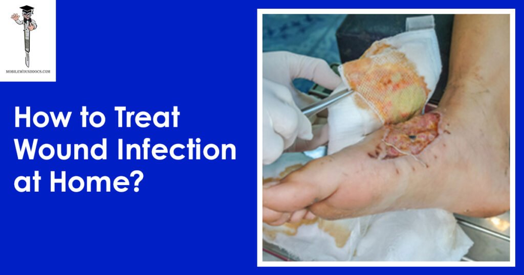
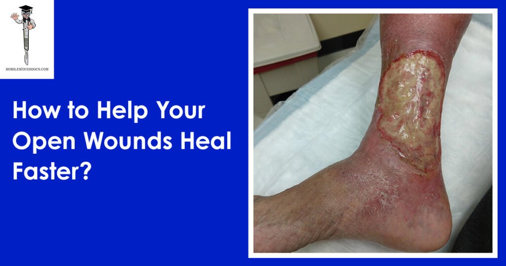
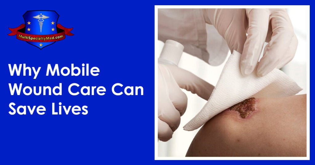
Daniel Davidson, MD, MBA, DBA, PHD Introduction: The physiological process of wound healing is intricate and includes several phases, such as tissue creation, remodeling, and inflammation. Most of the time, wounds move through these phases quickly, resulting in a successful closure and the preservation of tissue integrity. But occasionally, the healing process could take longer than expected, which could present difficulties for both patients and medical professionals. Numerous variables, from underlying medical issues to environmental circumstances, can cause delayed wound healing. We examine the reasons, signs, and available treatments for delayed wound healing in this article. What is Delayed Wound Healing? A condition known as “delayed wound healing” occurs when the body’s normal healing process—which normally involves mending damaged tissue—takes longer than anticipated or does not proceed through the phases in a reasonable amount of time. The biological process of wound healing is intricate and includes tissue creation, remodeling, and inflammation. In most cases, wounds close successfully and tissue integrity is restored as they move through these phases in a timely manner. Delays can occur from a variety of circumstances interfering with the natural healing process. These could include underlying medical disorders that affect blood flow, weaken the immune system, or obstruct the supply of vital nutrients needed for tissue regeneration, such diabetes, vascular disease, or malnutrition. Another frequent reason for delayed wound healing is infection, which is brought on by microbiological invasion, which can also worsen tissue regeneration, lengthen inflammation, and raise the risk of consequences. Causes of Delayed Wound Healing: A number of things can cause delayed wound healing, and all of them can make it more difficult for the body to restore injured tissue. These are a few typical reasons: Underlying Health Conditions: Wound healing can be severely hampered by long-term conditions such diabetes, vascular disease, autoimmune disorders, and malnutrition. These ailments may weaken the immune system, impair blood flow to the wound site, or obstruct vital nutrients needed for tissue healing. Infection: Infected wounds have a higher probability of taking longer to heal. Infections with bacteria, fungi, or viruses can exacerbate tissue regeneration, prolong inflammation, and raise the possibility of consequences including sepsis or abscess formation. Inadequate Blood Supply: To support tissue regeneration and healing at the wound site, enough blood flow is necessary to deliver nutrients and oxygen. Healing can be hampered by conditions that impair blood supply, such as venous insufficiency or peripheral artery disease. Drugs: A number of drugs have the potential to obstruct the body’s natural healing processes, delaying the healing of wounds. Corticosteroids, immunosuppressants, and nonsteroidal anti-inflammatory medications (NSAIDs) are a few examples. Smoking:  By narrowing blood vessels, lowering oxygen flow to tissues, and compromising immunological function, tobacco use can hinder the healing of wounds. Smokers are more likely to experience problems after surgery or an injury, including delayed wound healing. Advanced Age:  Aging is linked to alterations in the structure of the skin, a decrease in the generation of collagen, and a compromise in immunological function, all of which can cause delays in the healing of wounds. Nutritional Deficiencies: Encouraging the body’s healing processes requires a healthy diet. Wound healing can be hampered by deficiencies in protein, vitamins (especially C and A), and minerals (such iron and zinc). Obesity: Carrying too much weight around can put strain on wounds, obstruct blood flow, and raise the possibility of problems like infection. Poor outcomes after surgery or injury and delayed wound healing are linked to obesity. Poor Wound Care: Inadequate wound care can slow down the healing process and raise the risk of complications. This includes inappropriate cleaning, dressing selection, and failure to shield the site from additional trauma or infection. Symptoms of Delayed Wound Healing: Persistent Redness: Prolonged redness or inflammation in the vicinity of the wound may be a sign of continued inflammation and poor healing. Swelling: Prolonged edema or swelling close to the wound site may be a sign of a delayed healing process, which is frequently brought on by fluid buildup and poor lymphatic drainage. Warmth: If the skin around the incision feels warmer than the surrounding skin, there may be persistent inflammation and insufficient healing taking place. Pain: Continued or worsening pain at the site of the wound, especially during the initial phases of healing, may be a sign of underlying problems such tissue damage, nerve involvement, or infection. Increased Drainage: Extended periods of significant pus, blood, or clear fluid drainage from wounds may be a sign of infection or delayed healing. Reopening of the Wound: After first closure, wounds that frequently deteriorate or reopen may be a sign of underlying problems with tissue regeneration and healing. Development of Granulation Tissue: A symptom of the healing process, granulation tissue takes the form of pink or red tissue in the wound bed. On the other hand, extensive or protracted granulation tissue production could be a sign of postponed healing. Symptoms throughout the system:  Systemic symptoms like fever, chills, weariness, and malaise can appear in severe cases of delayed wound healing, especially if an infection is present. Treatment of Delayed Wound Healing: Wound Debridement: Objective: By minimizing the chance of infection and establishing a clean wound bed, removing dead or necrotic tissue from the wound site aids in the healing process. Techniques: There are a number of ways to accomplish debridement, including as mechanical, enzymatic, autolytic, or surgical approaches. Procedure: Depending on the features and severity of the wound, medical professionals will carefully remove non-viable tissue using sharp instruments, specialty dressings, or topical treatments. Infection Control: Objective: In order to lower inflammation, avoid systemic problems, and encourage tissue regeneration, it is essential to treat underlying infections. Method: Depending on the kind of infection present and the outcomes of culture and sensitivity testing, medical professionals may recommend antimicrobial therapy, such as antibiotics, antifungals, or antivirals. Monitoring: Treatment choices are based on a routine evaluation of the site for indicators of infection, such as elevated redness, edema, temperature, or purulent drainage. Optimizing Nutrition: Objective: By giving the body the vital

Scroll to Top