Daniel Davidson, MD, MBA, DBA, PHD
Introduction:
Decubitus ulcers, sometimes referred to as bedsores or pressure ulcers, are more than just superficial cuts; they are a result of persistent friction, shear, and pressure that gradually deteriorates the skin and underlying tissues. These ulcers usually progress through multiple phases of severity, each with unique traits and therapeutic and diagnostic consequences. Understanding the distinct phases is vital for precise evaluation, handling, and avoidance of decubitus ulcers.
Stage I:
Often seen in those with restricted movement or those confined to a bed or wheelchair, stage I decubitus ulcers are the first indications of skin damage brought on by continuous pressure. At this point, the skin feels warmer or colder than the surrounding tissue and may seem reddish or discolored. There are no obvious symptoms of breaks or open wounds on the skin despite the redness. On the other hand, the affected area could feel sensitive or uncomfortable to the touch. With timely treatment, stage I ulcers can be reversed. This includes optimizing skin care practices including keeping the skin clean, moisturized, and free from friction or excess moisture, as well as alleviating pressure by shifting postures frequently. To stop the progression to more severe phases, routine assessment and early intervention are crucial.
Stage Two:
The second stage of decubitus ulcers is characterized by partial-thickness skin loss, which means that the skin’s top layer (epidermis) and maybe its second layer (dermis) are affected. Usually manifesting as shallow open ulcers, these wounds can also look like superficial crater-like lesions, blisters, or abrasions on the skin’s surface. There’s a chance that the surrounding skin will be pink or red and swollen. Because the skin’s protective barrier has been compromised, even though the ulcer is not yet deep, it is more susceptible to infection than Stage I ulcers. When treating Stage II ulcers, it’s important to thoroughly clean the wound, remove any dead tissue, and use protective coverings to encourage healing and stop infection. To make sure the ulcer worsens, regular observation is necessary.
Stage Three:
Comparing stage III decubitus ulcers to previous stages, one can observe a notable advancement in tissue destruction. In Stage III, the ulcer penetrates the subcutaneous tissue below and spreads throughout the entire thickness of the skin. In contrast to the shallower wounds observed in Stages I and II, Stage III ulcers have a deeper degree of tissue involvement.
Stage III ulcer features consist of the following:
Deep ulceration:
The wound penetrates both the skin’s outer layer, the dermis, and the subcutaneous tissue below.
Visible necrosis:
The wound bed may contain necrotic tissue, such as slough, which is soft and yellow, or eschar, which is dry and black.
Undermining:
The surrounding tissue may be so damaged as to extend beneath the intact skin that encircles the wound.
Discoloration:
The ulcer’s surrounding skin may seem discolored, which is a sign of inflammation and tissue damage.
Potential exudate:
Depending on the degree of tissue damage and the existence of infection, stage III ulcers may produce drainage or exudate, which can vary in quantity and consistency.
Stage IV:
The most severe and advanced form of pressure ulcers is known as stage IV decubitus ulcers.
The tissue damage penetrates all skin layers, deep into the muscle, and occasionally even to the bone. These lesions are deep and wide, frequently resembling enormous craters with exposed necrotic (dead) tissue and surrounding healthy tissue being undermined. There can be fluid or discharge from the wound, and the surrounding skin might look discolored. In order to promote healing and avoid complications like infection or osteomyelitis (bone infection), treating Stage IV ulcers is difficult and usually calls for a multidisciplinary approach that includes aggressive wound care, debridement (removing dead tissue), infection control measures, and possibly surgical interventions like flap reconstruction or grafting.
Unstageable/Unclassified:
Decubitus ulcers defined as “unstageable” or “unclassified” occur when there is necrotic tissue covering the wound bed, making it impossible to precisely measure or stage the level of tissue destruction. The eschar or slough that usually covers these ulcers hides the depth of the lesion and makes it difficult for medical professionals to assess the entire amount of tissue involvement.
Eschar:
A hard crust of necrotic tissue that is dry, dark, or black that grows over the surface of a wound is called eschar. It can obstruct the evaluation of the underlying tissue and functions as a barrier to wound healing. Eschar can occur from tissue death brought on by pressure, ischemia, or other causes.
Slough:
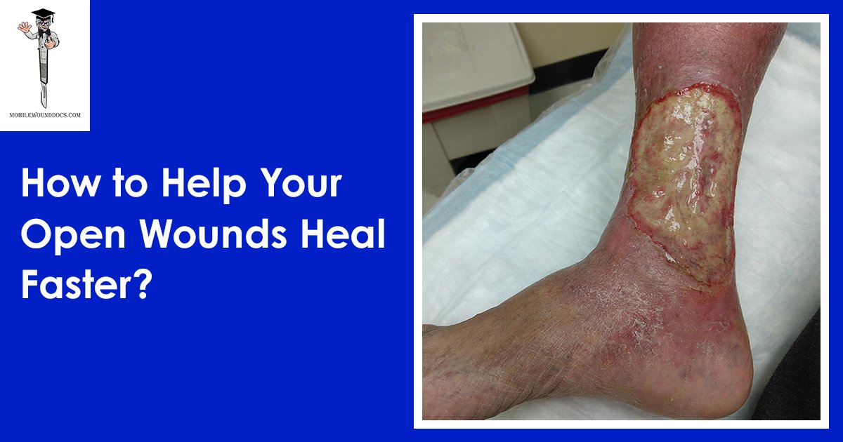
Slough is a type of moist, soft necrotic tissue that clings to the surface of wounds and appears yellow or white. It is made up of dead cells, debris, and exudate. By fostering a wet environment that is favorable to bacterial growth, it can impede the healing of wounds. Similar to eschar, slough can make it difficult to determine the actual depth of the incision and the degree of tissue damage.
The development of eschar or slough in decubitus ulcers that are not stageable or classed makes it challenging to assess if deeper tissues are affected by the lesion or if it has reached the full thickness of the tissue. Because of this, these ulcers cannot be classified using conventional staging criteria, which designates a certain severity level.
Healthcare professionals usually concentrate on wound debridement, which is removing necrotic tissue to reveal the underlying healthy tissue and promote wound healing, in order to treat ulcers that cannot be staged or classed. Depending on the features of the wound and the patient’s general state, debridement treatments might be mechanical, surgical, enzymatic, or autolytic.
Healthcare professionals are able to precisely stage the decubitus ulcer and create a suitable treatment strategy once the wound bed has been sufficiently debrided and the degree of tissue destruction is apparent. This could entail treating underlying risk factors to stop recurrence as well as promoting wound healing using dressings, topical therapies, pressure redistribution, and nutritional assistance.
It is crucial to regularly examine and reevaluate the treatment strategy in order to follow the healing process of wounds. Healthcare professionals can improve patient outcomes and lower the risk of complications by addressing decubitus ulcers that cannot be staged or categorized.
Conclusion:
Decubitus ulcers develop in different levels of severity, each with its own clinical characteristics and therapeutic consequences. In order to stop the disease from progressing to more severe stages and lower the chance of consequences, early detection and treatment are essential. In order to accelerate wound healing and enhance patient outcomes, healthcare providers need to do routine assessments and use the proper therapies.


